This was followed shortly by the now well-known initial trabecular bone microstructure article. Goldstein would name the technique ‘microcomputed tomography’, and this collaboration led to the first publication of microCT analysis of bone architecture, an evaluation of subchondral bone in experimental osteoarthritis. That same year, through connections at Henry Ford Hospital, Feldkamp was introduced to Steven Goldstein, an orthopedic biomechanician at the University of Michigan. A serendipitous meeting between Feldkamp and Michael Kleerekoper of Henry Ford Hospital led to the first scan of bone tissue, an iliac crest biopsy, and resulted in the first public evidence of microCT: an abstract from the 1983 meeting of the American Society for Bone and Mineral Research. He then developed the cone-beam algorithm to reconstruct fully 3D images from those projections. Expanding on the concepts of clinical CT systems, Feldkamp conceived of using a cone-beam x-ray source and 2D detector and rotating the sample itself through 360°. In the early 1980s, Ford Motor Company physicist Lee Feldkamp developed the first microCT system to evaluate structural defects of ceramic automotive materials. In 1979, Allan Cormack and Godfrey Hounsfield were awarded the Nobel Prize in Physiology or Medicine for the development of computer-assisted tomography and, by the late 1970s, clinical computed tomography (CT) was in widespread use however, these systems were limited in resolution and yielded only 2D reconstructions as they relied on line x-rays and linear array detectors.

The attenuation therefore depends on both the sample material and source energy and can be used to quantify the density of the tissues being imaged when the reduced intensity beams are collected by a detector array. As an x-ray passes through tissue, the intensity of the incident x-ray beam is diminished according to the equation, I x = I 0e −μx, where I 0 is the intensity of the incident beam, x is the distance from the source, I x is the intensity of the beam at distance x from the source, and μ is the linear attenuation coefficient.

The principle of microCT is based on the attenuation of x-rays passing through the object or sample being imaged. A three-dimensional rendering of the sample is achieved by scanning at different angles of rotation and reconstructing through transformation of two-dimensional projections. The radiation is attenuated by the sample, and this attenuation is measured by a charge-coupled device (CCD) camera with a phospholayer coating to convert x-rays to visible light. A micro-focus x-ray tube, or synchrotron emitter for monochromatic beam generation, produces radiation, which is collimated and passed through the object. Principal components of a microcomputed tomography scanner. This non-destructive imaging modality can produce 3D images and 2D maps with voxels approaching 1 μm, giving it superior resolution to other techniques such as ultrasound and magnetic resonance imaging (MRI).
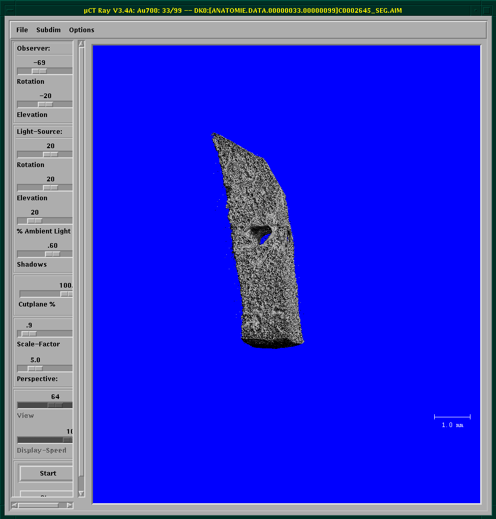
#Microct video rotate 3d reconstructed image series
Reconstruction of a 3D image is performed by rotating either the sample (for desktop systems) or the emitter and detector (for live animal imaging) to generate a series of 2D projections that will be transformed to a 3D representation by using a digital process called back-projection. MicroCT equipment is composed of several major components: x-ray tube, radiation filter and collimator (which focuses the beam geometry to either a fan- or cone-beam projection), specimen stand, and phosphor-detector/charge-coupled device camera (Figure 1). Microcomputed tomography (microCT or μCT) is a non-destructive imaging tool for the production of high-resolution three-dimensional (3D) images composed of two-dimensional (2D) trans-axial projections, or ‘slices’, of a target specimen.


 0 kommentar(er)
